Presenter: Andrea Costanza, DO, Nanda Channaiah, DO, Kevin Belasco, DO, Kevin Dehart, DO, Aaron Bruce, DO and Roger Sica, DO
Dermatology Program: NOVA Southeastern University – Suncoast Hospital
Program Director: Richard Miller DO, FAOCD
Submitted on: January 30, 2007
CHIEF COMPLAINT: Adolescent-onset rash and progressively worsening symptoms
CLINICAL HISTORY: We present a 25 y/o female with a history of adolescent-onset rash and progressively worsening symptoms. Upon review of history, the patient admitted to recurrent episodes of headaches, fainting spells, flushing, pruritus, palpitations, wheezing, abdominal pain, and vomiting within the last year. Her skin lesions periodically become raised, erythematous, and pruritic, which are exacerbated with “asthma attacks.” Exercise and Naprosyn worsen her symptoms and induce acute attacks. Neurocardiogenic syncope was also noted in medical history.
Previous Treatment: Laser, intralesional and topical corticosteroids.
Other information: Medications upon presentation include Advair, Combivent, Synthroid, Singulair, and Yasmine.
PHYSICAL EXAM:
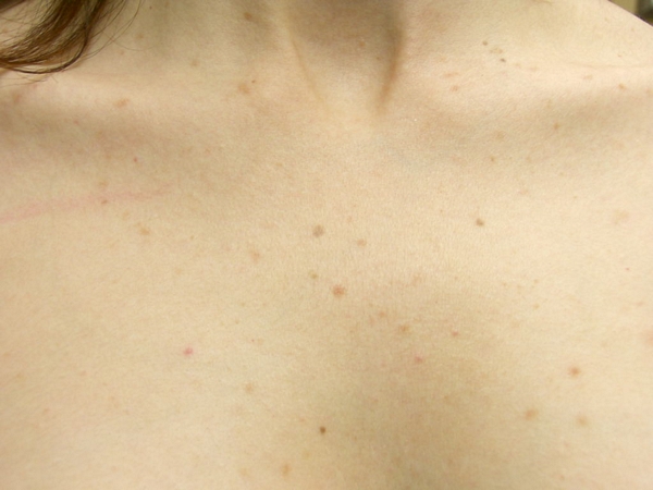
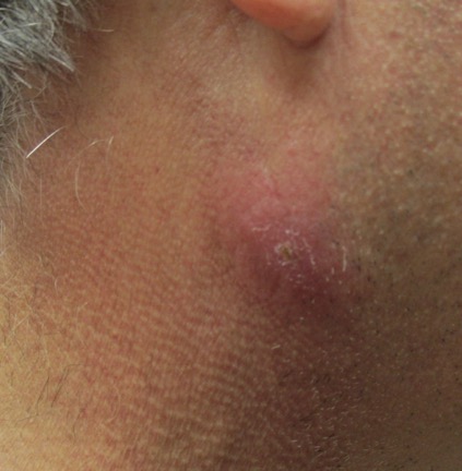
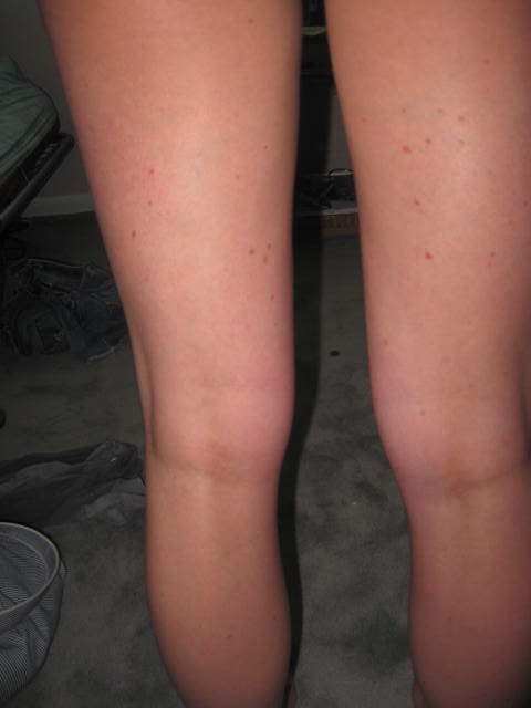
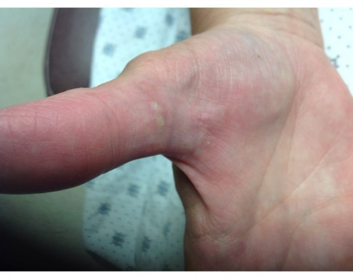
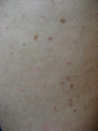
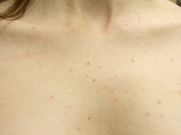
Examination reveals numerous reddish-brown, well-demarcated macules and papules less than 1 cm in diameter. These lesions are symmetrically distributed, most numerous on the trunk, buttocks, and proximal extremities. The face is spared.
LABORATORY TESTS:
CBC, CMP, rheumatoid factor, ANA, SPEP, and serum tryptase were all within normal limits. In addition, karyotype and bone marrow biopsy with flow cytometry was also within normal limits. Peripheral blood smear showed the pelger-huet anomaly.
DERMATOHISTOPATHOLOGY:
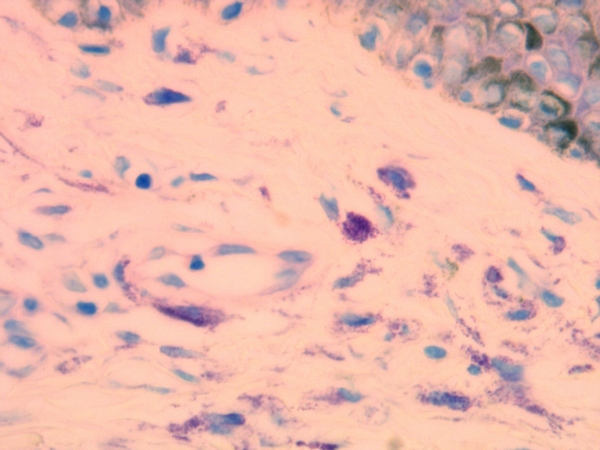
Increased superficial dermal perivascular cells with granular cytoplasm.

The cells stained metachromatically with Giemsa.
DIFFERENTIAL DIAGNOSIS:
1. Systemic Mastocytosis
2. Carcinoid syndrome
3. VIPoma
4. Zollinger-Ellison syndrome
5. Pheochromocytoma

