Presenter: Keoni Nguyen, DO; Dawn Sammons, DO; Ramona Nixon, DO; Shannon Campbell, DO
Dermatology Program: Ohio University COM/ O’Bleness Memorial Hospital
Program Director: John P. Hibler, DO, FAOCD
Submitted on: May 28, 2008
CHIEF COMPLAINT: Irritation to the bilateral forearms, hands, neck, and face
CLINICAL HISTORY: A 38-year-old Caucasian male presented to our office with a one-year history of chronic blisters and non-healing ulcers on both of his upper extremities. His neck and face would incur a pruritic rash with prolong exposure to the sun. His symptoms are worse in the summer. The patient was previously treated with oral prednisone for ten days and an unknown topical cream; neither of which alleviated his symptoms. He denies any fevers, chills, or general myalgias. He reports a history of warts and seasonal allergies. He also endorses consuming 4-5 beers per night and 12 on the weekends and tobacco use of 1 pack per day. He works as an electrician. He had been to several countries outside of the U.S. in the past, while in the military. Denies any allergies to medications.
PHYSICAL EXAM:
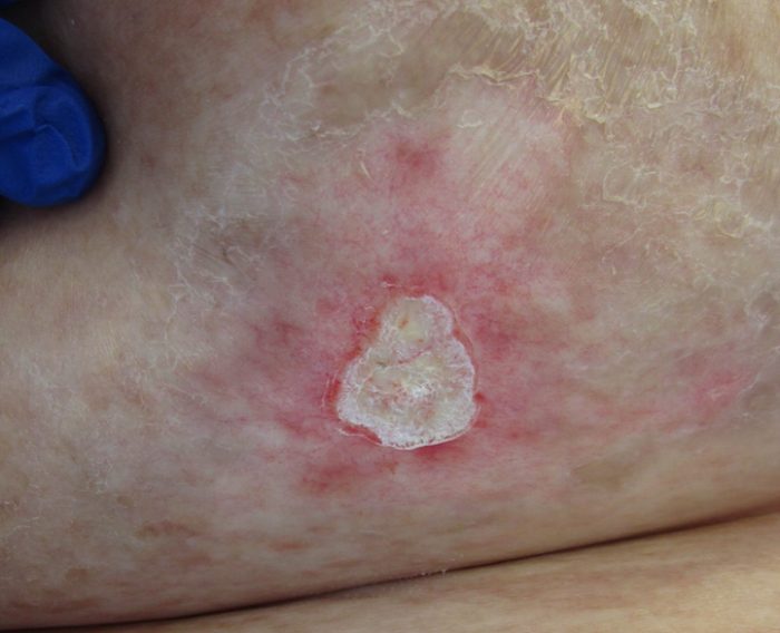
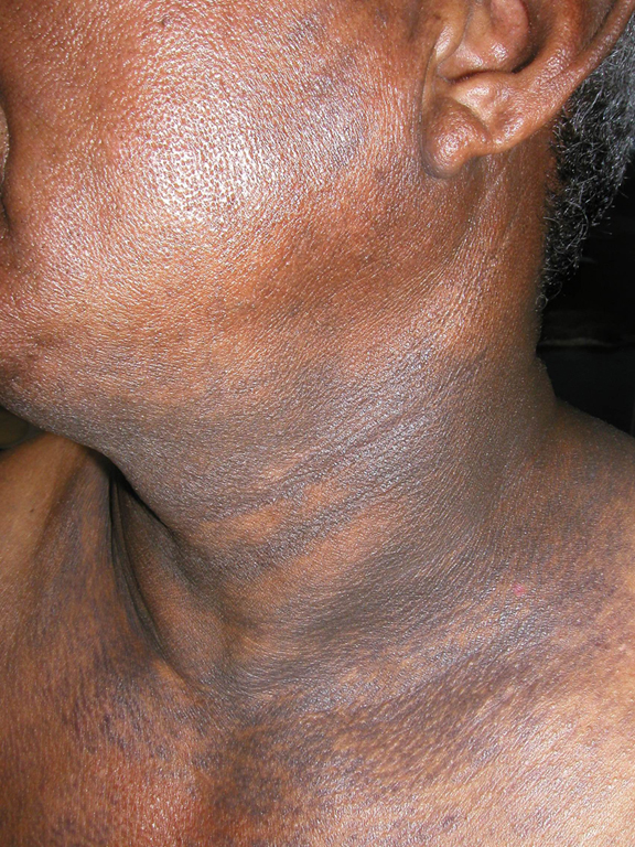
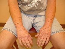
On examination, the patient presented with malar hypertrichosis. The neck region had areas of sclerodermatous thickenings. The dorsal hands bilaterally revealed multiple ulcerations and erosions. On closer examination of his dorsal hands, they were studded with milia.
LABORATORY TESTS:
Initial labs: Porphyrins, 24 hour UA: Uroporphyrin 307, Heptacarboxylate porphyrin 249, Corproporphyrin 95; Porphyrins, fecal: Uroporphyrin 34 nmol/g, Protoporphyrin 70 nmol/g, Porphyrin normal; CBC –wnl; LFT’s: ALT 151, AST 123; Hepatitis serology: Hepatitis C AB – Reactive, Hep A IgM – Nonreactive, Hep B CORE-M AB – Nonreactive, Hep B SURF AG – Nonreactive.
Follow-up labs/tests: HIV1-HIV-2 AB Nonreactive; Ferritin 659 ng/mL, G6PD 14.1, Hep C RNA Quant PCR 6.2; HFE Molecular Analysis for Hereditary Hemochromatosis Genotype: Exon 4 C282Y Locus – Wild type/ No mutation, Exon 2 H63D Locus – Heterozygous mutation (1 abnl allele), Exon 2S65C Locus – Wild type/ No mutation.
DERMATOHISTOPATHOLOGY:
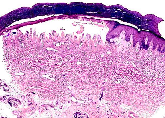
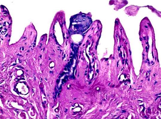
Two 4 mm punch biopsies were performed. The first biopsy (specimen A) was on the right lateral forearm for hematoxylin-eosin (H&E) staining. The second biopsy (specimen B) was on the right medial forearm for Direct Immunofluorescence (DIF).
Specimen A: The biopsy revealed hyperkeratosis, parakeratosis, and a subepidermal blister. A perivascular chronic inflammatory infiltrate admixed with eosinophils was present in the superficial and deep dermis and focally extended into the subcutis. Alcian blue/PAS staining showed thickened basement membranes and around blood vessels in the superficial dermis.
Specimen B: Direct immunofluorescence showed trace +IgM, C3, and C5b-9 at the dermo-epidermal junction in a granular pattern and in blood vessel walls in the superficial dermis. DIF for albumin, IgG, and IgA was negative.
DIFFERENTIAL DIAGNOSIS:
1. Polymorphic light eruption
2. Pseudoporphyria
3. Epidermolysis bullosa
4. Porphyria Cutaneous Tarda
5. Phototoxic and bullous drug eruptions


