Presenter: Susun Bellew, DO; Grace K. Kim, DO; Saira Momin, DO; Brent Michaels DO
Dermatology Program: Valley Hospital Medical Center, Las Vegas, Nevada
Program Director: James Q. Del Rosso, D.O.
Submitted on: January 30, 2010
CHIEF COMPLAINT: “rash” on lower extremities
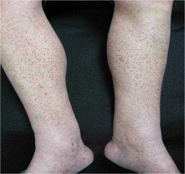
CLINICALHISTORY: A 62-year-old Italian man presented to the office for evaluation of a “rash” that started on his lower extremities. The patient reports a persistent eruption for 4 years that originally started on his lower extremities which progressed to his arm, palms, and low back. He also reported occasional pruritus of the lesions which was not severe. The patient and his wife both denied any suspected precipitating factors. He also denied constitutional symptoms such as fever, chills, weight loss, myalgia, or arthralgia. The lesions were previously treated with topical triamcinolone 0.1% cream with no clinical improvement. Past medical history was significant only for Parkinson’s disease. There was no known history of other major medical disorders, such as metabolic diseases or malignancies. He also denied recent travel or history of any sexually transmitted diseases, including syphilis. No other family members or close personal contacts were afflicted with similar lesions.
PHYSICAL EXAM:
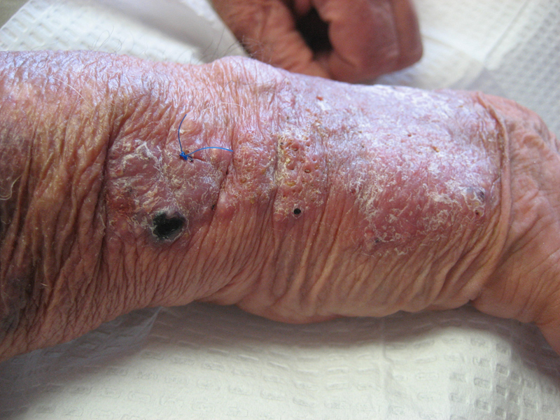
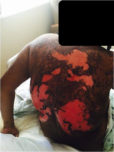
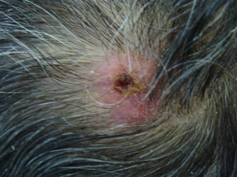
Examination revealed numerous symmetric 1-5mm red-brown scaly, focally keratotic papules on his lower legs and feet. Other anatomic regions of involvement included the arms, palms, abdomen, and back. Importantly, the lesions were monomorphic in appearance overall. No excoriations, vesicles, pustules, or burrows were noted. The face, neck, and scalp were spared, and no lesions were noted on his genitalia. Removal of the scales resulted in pinpoint bleeding. Oral mucosa, hair, and nails were not affected. There was no cervical, axillary, or inguinal lymphadenopathy.

LABORATORY TESTS: N/A
DERMATOHISTOPATHOLOGY:
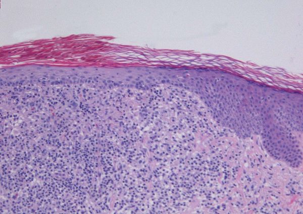
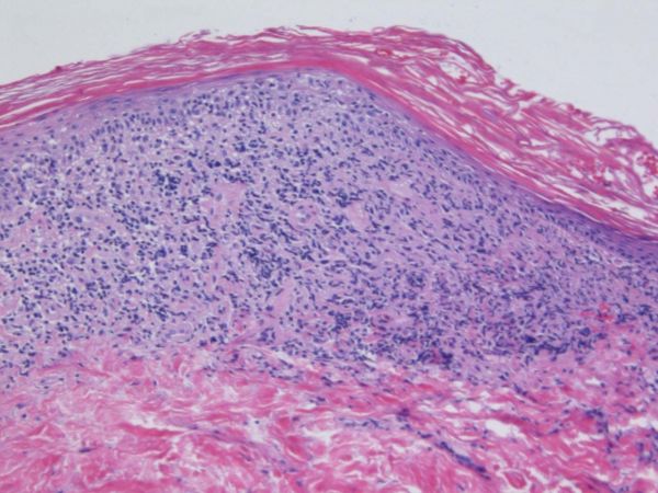
A 4mm punch biopsy was obtained from one of the lesions and sent for histological analysis. Histopathologic examination revealed a band-like infiltrate of lymphocytes and histiocytes. There was also exocytosis of lymphocytes into the overlying epidermis, with mild spongiosis with focal vacuolar alteration of the basal layer keratinocytes. In addition, there was an absence of the granular layer with overlying compact orthokeratosis and focal parakeratosis. Epidermal thinning with flattened rete ridges were also observed. The adjacent epidermis revealed the preservation of granular layer with overlying basket-weave orthokeratosis.
DIFFERENTIAL DIAGNOSIS:
1. Acrokeratosis verruciformis of Hopf
2. Actinic keratosis
3. Hyperkeratosis lenticularis perstans (Flegel’s disease)
4. Hyperkeratosis follicularis et parafollicularis in cutem penetrans (Kyrle’s disease)
5. Keratosis follicularis (Darier’s disease)


