Presenter: Chelsea Lee, DO, Payal Patel, DO, Kimball Silverton, DO
Dermatology Program: Genesys Regional Medical Center
Program Director: Kimball Silverton, DO
Submitted on: March 25, 2012
CHIEF COMPLAINT: rash located over her back, chest, neck, face, upper and lower extremities for two weeks
CLINICAL HISTORY: A 71-year-old woman presented to the dermatology clinic with a rash located over her back, chest, neck, face, upper and lower extremities for the duration of two weeks. The symptoms included stinging and pruritus. Her past medical history was significant for hypertension and GERD. She had never experienced a rash similar to this in the past, and she denied any recent changes in her health or lengthy exposure to sunlight. In addition, she denied any fevers, joint pains, a history of skin disease, or photosensitivity. The patient did, however, state that she began terbinafine for the treatment of onychomycosis two weeks prior to the development of the rash.
PHYSICAL EXAM:
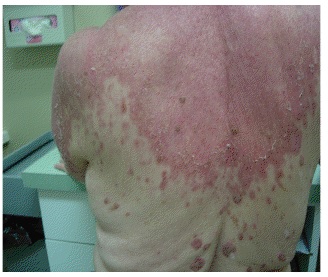
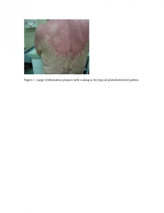
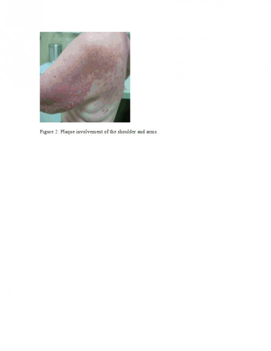
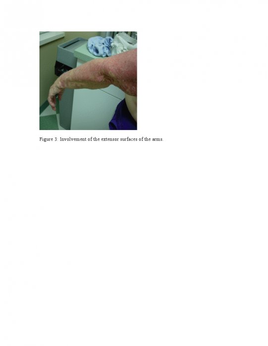
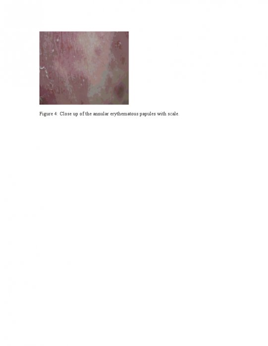
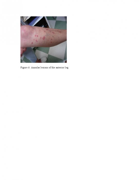
On physical exam, the patient had large erythematous plaques with scaling in a photodistributed pattern, including her upper back, upper chest, and extensor surfaces of the arms, with sparing of the underarms. In addition, multiple annular erythematous papules and plaques were found scattered around the periphery of the large plaques. There was marked erythema of her face and neck, which was more pronounced on her forehead and cheeks. Multiple annular, scaly, erythematous papules and plaques were seen on the anterior surface of the legs.

LABORATORY TESTS:
Laboratory results indicated a high ANA titer with a homogenous ANA pattern. Anti-Ro (SS-A) and anti-La (SS-B) were significantly elevated. Negative antibodies included anti-histone, anti-dsDNA, anti-ssDNA, anti-Scl-70, and anti-Smith. Of note, her glucose and ALT were also mildly elevated.
DERMATOHISTOPATHOLOGY:
A 3mm punch biopsy revealed orthokeratosis and parakeratosis with effacement of the rete ridges in the epidermis. Vacuolar alteration of basal keratinocytes, and a few necrotic keratinocytes were present as well in the epidermis. A lymphocytic predominant infiltrate was seen in a superficial and mid-dermal perivascular, interstitial, and peri-eccrine pattern. A diagnosis of vacuolar interface dermatitis was made.
DIFFERENTIAL DIAGNOSIS:
1. Connective Tissue Disease
2. Interface Drug Reaction
3. Photoeruption
4. Drug-Induced SCLE

