Presenter: Christina Feser DO, Arathi Goldsmith DO, Peter Saitta DO, Jean Holland DO, Stephen Olsen DO
Dermatology Program: Oakwood Southshore Medical Center
Program Director: Dr. Steven Grekin
Submitted on: August 5, 2012
CHIEF COMPLAINT: Resistant rash
CLINICAL HISTORY: September 2009-May 2010: A 61-year-old Caucasian female presented to our clinic with a 1-year history of a waxing and waning eruption located on the trunk and extremities. The patient denied pain and pruritus. No significant past medical history or family history was noted. The patient was otherwise healthy. A review of systems was unremarkable. May 2010-November 2010: The rash failed to resolve after multiple courses of topical and systemic steroids. Despite treatment, the eruption generalized. November 2010-August 2012: The patient’s condition continued to evolve becoming more diffuse throughout the trunk and extremities despite additional treatment trials with dapsone and acitretin. Finally, cyclosporine was added leading to the diagnostic eruption.
PHYSICAL EXAM:
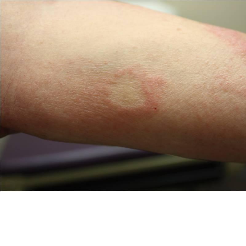
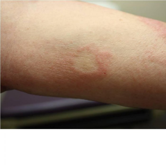
September 2009-May 2010: The eruption was characterized by annular, edematous to eczematous plaques on the extremities, giving the illusion
of an urticarial eruption.
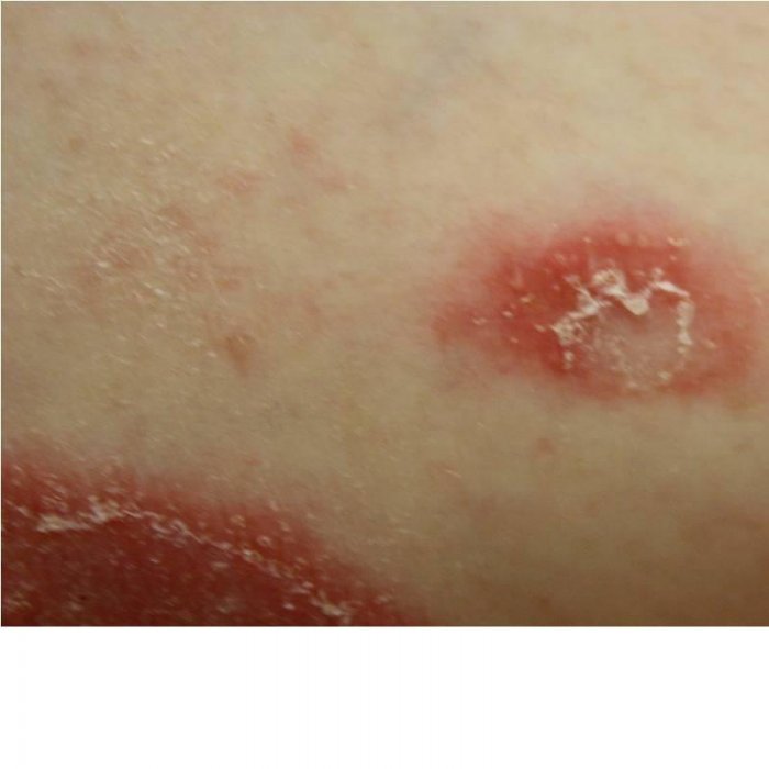
May 2010-November 2010: The patient noted to have an appearance of erythematous annular to arcuate plaques studded with small, tense pustules, preferentially involving the flexure surfaces bilaterally. Some plaques collided forming polycyclic lesions with serpiginous borders. A fine trailing scale was prominent, reminiscent of an erythema annulare centrifugum presentation.
November 2010-present: The patients lesions generalized and became beefy red in color coalescing to form juicy plaques. The minute pustule, the trailing scale, and annular configuration were no longer apparent.

LABORATORY TESTS:
No significant abnormalities were noted in complete blood count with differential, complete metabolic profile, iron studies, complement levels, thyroid studies, lipid panel, and hemoglobin A1C. The Hepatitis panel was non-reactive. Serum protein electrophoresis was negative. Respiratory allergen profile reactive only to dog dander. Chest X-ray with no acute disease.
DERMATOHISTOPATHOLOGY:
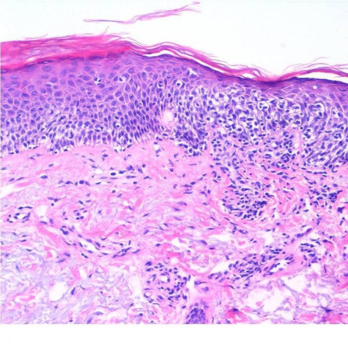
H&E September 2009-May 2010:
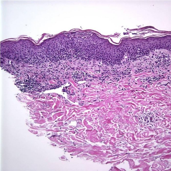
H&E May 2010-November 2010:

H&E November 2010-present:
DIFFERENTIAL DIAGNOSIS:
1. Allergic Contact Dermatitis
2. Atopic Dermatitis
3. Cutaneous T-Cell Lymphoma
4. Sneddon-Wilkinson Disease

