Presenter: Douglas M Richley, DO
Dermatology Program: Northeast Regional Medical Center
Program Director: Lloyd J Cleaver, DO
Submitted on: January 15, 2015
CHIEF COMPLAINT: Initial evaluation of a generalized, pruritic eruption
CLINICAL HISTORY: A 44-year-old obese Caucasian male presented for initial evaluation of a generalized, pruritic eruption. The eruption had been present for approximately three weeks, beginning on his elbows and subsequently spread to involve his back, abdomen, knees, and posterior thighs. The patient denied recent illness or changes in medications and also admitted to having frequent bouts of blurry vision over the past couple of weeks. No previous treatments.
PHYSICAL EXAM:
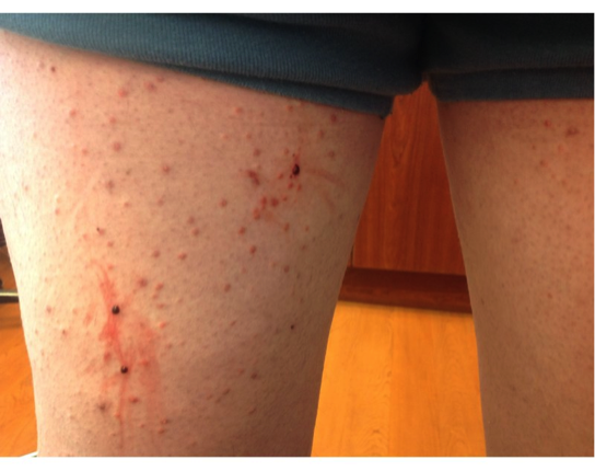
On physical examination, there were multiple excoriated, orange-red firm papules on an erythematous base. Lesions were distributed on the abdomen, back, bilateral elbows, knees, and posterior thighs. The remainder of the dermatologic exam was normal. A scabies prep was performed which showed no mites, ova, or scybala.

LABORATORY TESTS:
Baseline labs were also ordered which included complete blood count (CBC), complete metabolic panel (CMP), fasting lipid panel, amylase, lipase, hemoglobin A1C, and thyroid-stimulating hormone (TSH). The results of the blood work came back with the following remarkable results: triglycerides 4931 mg/dL, total cholesterol 410 mg/dL, hemoglobin A1C 12% (avg. glucose – 298 mg/dL), amylase normal, lipase normal.
DERMATOHISTOPATHOLOGY:
A 4mm punch biopsy of one of the papules was performed. The lesion was sent for histologic examination. On histology, hematoxylin and eosin (H & E) stained sections demonstrated foamy, xanthomatous histiocytes scattered throughout multiple levels of the dermis, being most concentrated in the superficial dermis. These histologic findings are consistent with xanthoma.
DIFFERENTIAL DIAGNOSIS:
1. Eruptive Xanthoma
2. Dermatitis Herpetiformis
3. Lichen Planus
4. Granuloma Annulare
5. Arthropod Assault


