Presenter: Carmen A Julian DO, Irina Milman DO, Eugene Sanik DO
Dermatology Program: PCOM/North Fulton Hospital Medical Campus
Program Director: Marcus B Goodman, DO FAOCD
Submitted on: August 3, 2015
CHIEF COMPLAINT: Asymptomatic rash with purplish discoloration on her trunk, extremities and ears
CLINICAL HISTORY: A 40 year old female presenting with fever, cough, hemoptysis and an asymptomatic rash with purplish discoloration on her trunk, extremities and ears. The patient reported the rash started 5 days prior while she was undergoing inpatient treatment for pneumonia at a nearby hospital. The rash started on day two of her admission. She denies pain, bleeding or pruritus associated with the involved areas. She also denied any constitutional symptoms. She received empiric intravenous antibiotics for community-acquired pneumonia on her previous admission. She states that a punch biopsy was performed at her recent outside admission, but our attempts to obtain a report were unsuccessful. No specific treatment for her rash had yet been implemented. The patient left AMA from the previous hospital and presented to our hospital with worsening symptoms. Prior medical records, including the biopsy and antibiotic treatment, again were unavailable. She does admit to tobacco abuse and cocaine abuse, most recently 11 days prior during Independence Day weekend. She denies intravenous drug use.
PHYSICAL EXAM:
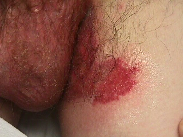
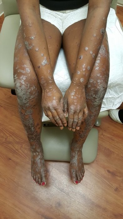
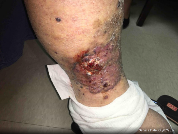
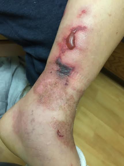
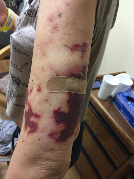
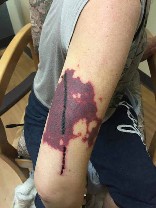
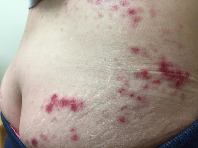
On general examination, the patient was non-toxic, afebrile, with stable vital signs. Skin examination revealed tender, violaceous macules and patches on the trunk, extremities, and ears. There were areas of retiform, stellate purpura progressing to bullae on the upper arms and lower legs. Additionally, there was a pink-to-erythematous macular-papular eruption on the entire back. A small eschar was seen on the left medial ankle. No desquamation or mucosal involvement was noted. On the left outer arm, there was a healing, sutured punch biopsy wound.

LABORATORY TESTS:
White count 7.02, Hemoglobin 10.7, Platelets 535, C-reactive protein 2.85, lactic acid 1.70. The basic metabolic panel was within normal limits. Chest x-ray and CT-Angiography chest showed left lower lobe, right upper lobe opacities, splenomegaly, and bilateral axillary lymphadenopathy. HIV- non-reactive, ANA- negative, P-ANCA and C-ANCA titers all <1
DERMATOHISTOPATHOLOGY:
According to the patient, a punch biopsy was performed at her recent admission to an outside hospital, and a biopsy scar is noted on clinical examination. Multiple attempts to obtain the report were unsuccessful and the outside hospital denied having any records matching the name and date of birth of our patient. Typically biopsies of direct areas of involvement of this condition, however, would reveal a leukocytoclastic vasculitis with varying involvement of superficial and deep dermal vessels.
DIFFERENTIAL DIAGNOSIS:
1. Wegener’s granulomatosis
2. Cryoglobulinemia
3. Cocaine levamisole toxicity
4. Septic Vasculitis
5. Polyarteritis nodosa


