Presenter: Shannon McKeen, DO
Dermatology Program: MSUCOM/Lakeland Regional Medical Center
Program Director: Mark A. Kuriata, DO, FAOCD
Submitted on: October 22, 2016
CHIEF COMPLAINT: Persistent rash on the right breast
CLINICAL HISTORY: A 63-year-old Caucasian female with a persistent rash on the right breast. In February of 2000, the patient underwent excision of ductal carcinoma in situ, a high nuclear grade with focal micro-invasion of the right breast. This was followed by eight weeks of radiation therapy and a five-year course of tamoxifen. She did not seek to follow up imaging until 2004, which had shown calcifications. Biopsy of the calcifications showed recurrent ductal carcinoma in situ. At that time, the surgeon recommended mastectomy with axillary lymph node dissection. The patient refused due to concerns over loss of function and swelling in the arm. In 2016, several months prior to her presentation in our office, the patient developed a rash on the right breast. The rash involved the areola and periareolar skin. The patient described the rash as red, itchy, and mild in severity. She was seen by her Gynecologist who gave her a topical corticosteroid cream, which helped improve the rash somewhat, only to return upon discontinuation. The patient also reported using a “diaper rash cream” which mildly improved her symptoms. The Gynecologist ordered a 3D mammography and subsequent ultrasound. Both reports were read as having dystrophic calcifications, recommending six months follow up exams. The patient was referred to our office for her persistent rash. At the initial consultation, we ordered an MRI of the right breast, referred her to the general surgeon who performed the initial excision in 2000, and the patient was given samples of flurandrenolide 0.05% cream to apply twice daily until follow up. The patient called several days later and refused MRI, as well as canceled the appointment with the general surgeon. She was instructed to return to the office for a biopsy.
PHYSICAL EXAM:
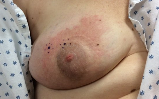
On the right breast, there was a fairly well-demarcated erythematous plaque with overlying scale and focal areas of induration upon palpation affecting the periareolar skin and areola. The right nipple was erythematous and everted. Cobblestone appearance of the superior aspect of the right areola noted. Well healed surgical incision site on the superior periareolar skin from prior surgery.
LABORATORY TESTS:
3D mammography in August 2016 showed a density with dystrophic calcification in the right subareolar and right upper outer breast deemed as benign-appearing and a six- month follow up was advised. A follow-up ultrasound one week later showed an 8 x 6 x 6 mm cystic structure in the immediate right subareolar region with dystrophic calcification deemed as benign-appearing and a six-month follow up was advised. The patient did not obtain the MRI ordered by our office.
DERMATOHISTOPATHOLOGY:
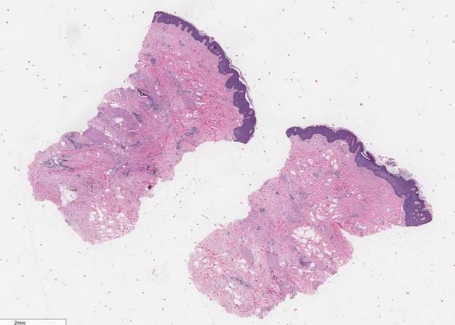
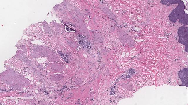
Two punch biopsies were obtained demonstrating an uninvolved epidermis and islands of atypical cells with cytologic pleomorphism and mitotic figures throughout the dermis with perivascular invasion. The cells stained positively for cytokeratin-7 and negative for cytokeratin-20.
DIFFERENTIAL DIAGNOSIS:
1. Mammary Paget’s Disease
2. Metastatic Breast Carcinoma
3. Candida Infection of the Breast
4. Allergic Contact Dermatitis of the Breast
5. Localized Atopic Dermatitis of the Breast


